Blood Work Education
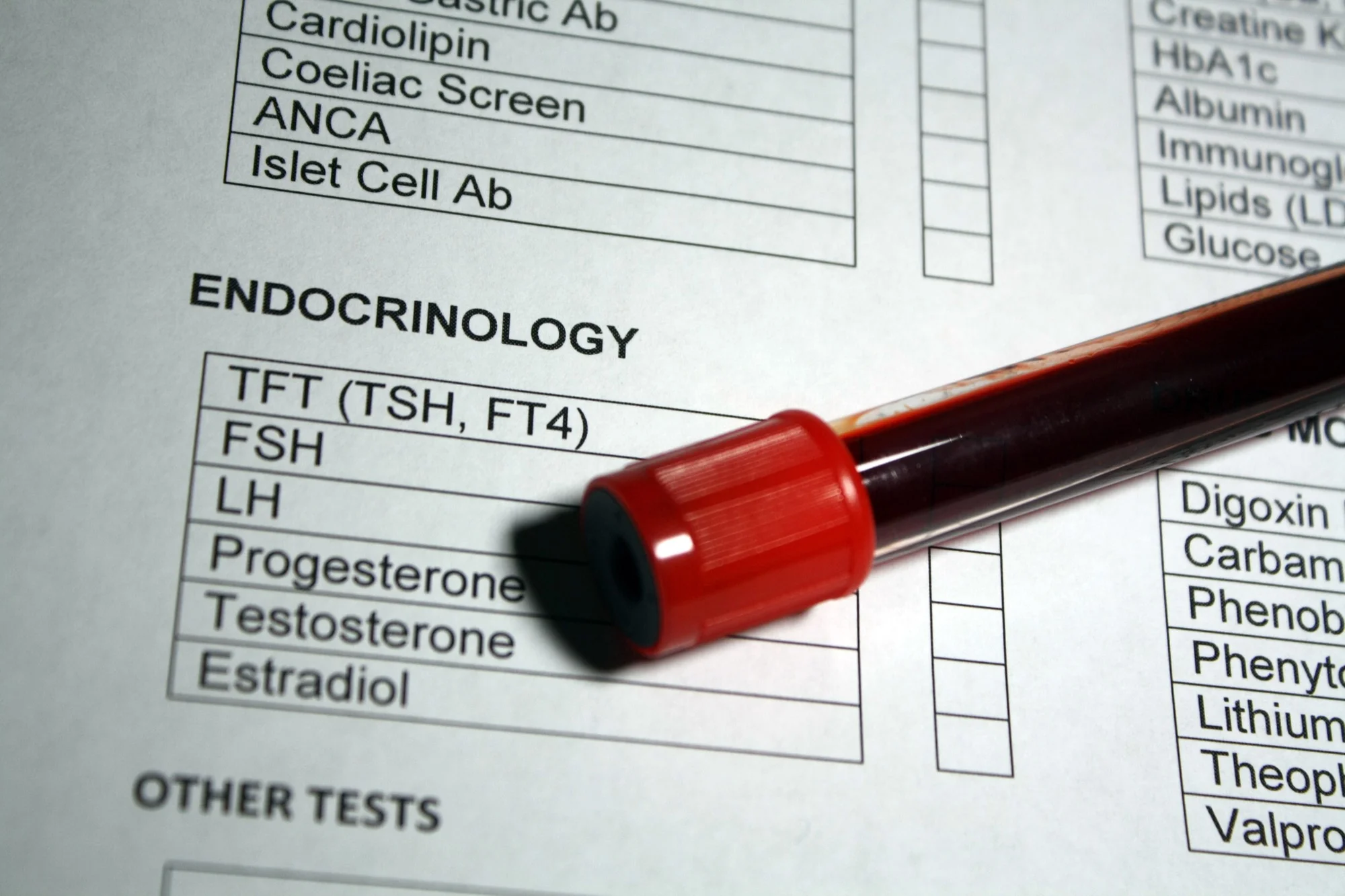
HORMONES & ENDOCRINE
Understanding your hormone panel -- testosterone, estradiol, SHBG, DHEA-S, FSH, LH, and PSA explained in plain language with clinical context.
Medically reviewed by Missy Zammichieli, DNP, APRN, FNP-BC · Updated March 2, 2026
THE BOTTOM LINE
- • Your hormone panel looks at testosterone, estrogen, and the signals that control them
- • Total testosterone alone doesn't tell the full story -- free T and SHBG matter just as much
- • Hormones are affected by sleep, body composition, stress, and metabolic health
- • Morning blood draws are essential for accurate testosterone results
INTRODUCTION
Hormones are chemical messengers that regulate energy, body composition, mood, sex drive, bone density, and cardiovascular health. When they're in balance, things run smoothly. When they shift -- from aging, stress, metabolic changes, or other factors -- the effects can show up everywhere.
A common misconception is that "checking hormones" means checking a single testosterone number. In reality, one number tells you very little. Your total testosterone can look normal while the amount your body can actually use is low. That's why this panel tests multiple markers that work together as a system.
Here's a simple way to think about how your hormones are regulated. Your brain tells your body how much testosterone to make, and your body reports back. Think of it like a thermostat -- the brain sets the target, the body produces, and signals loop back to keep things in range. Disruptions anywhere in that loop -- brain, pituitary gland, or gonads -- create different patterns on lab work. That's why testing multiple markers at once matters.
YOUR BIOMARKERS EXPLAINED
1. Testosterone, Total
This is the headline number -- your overall testosterone level. It measures all the testosterone in your blood: the portion bound to proteins and the small fraction floating freely.
Why it matters: Testosterone supports muscle mass, bone density, red blood cell production, fat distribution, libido, erectile function, mood, and cognitive function. In women, it contributes to libido, bone health, and muscle maintenance at much lower levels.
What the guidelines say: The American Urological Association defines testosterone deficiency as total T below 300 ng/dL, measured on at least two separate morning draws. The Endocrine Society uses a similar threshold. Most labs place the normal range between 264 and 916 ng/dL.
The aging trend: Testosterone declines about 1-2% per year after age 40 on average. But this isn't destiny. Some men maintain strong levels into their 70s and 80s. Body composition, sleep, and metabolic health play a huge role.
Common causes of low total T:
- • Obesity (body fat converts testosterone to estrogen)
- • Sleep deprivation
- • Chronic stress
- • Opioid use
- • Heavy alcohol use
- • Liver disease
- • Pituitary disorders
Women: Normal total testosterone in premenopausal women is typically 15 to 70 ng/dL.
2. Testosterone, Free
This is the testosterone your body can actually use. Only about 1-3% of your total testosterone circulates freely -- unbound to any protein -- and this is the fraction that enters cells and does the work.
Why it matters: You can have a total testosterone of 450 ng/dL (solidly normal) but if your SHBG is elevated, your free T may be low enough to cause symptoms. The reverse is also true -- borderline-low total T with low SHBG can mean your free T is perfectly adequate. Free T often gives a more accurate picture than total T alone.
When it's checked: The Endocrine Society recommends measuring free T when total T is near the lower boundary of normal, or when conditions that alter SHBG are present (obesity, aging, diabetes, thyroid disease, liver disease). Values below 5 to 9 ng/dL in men are commonly considered low.
Bioavailable testosterone is a related measure that includes free T plus a loosely-bound fraction that can also become active in tissues. It's sometimes reported alongside free T to give a fuller picture, especially when SHBG is significantly elevated.
3. Sex Hormone Binding Globulin (SHBG)
This protein locks up testosterone so your body can't use it. SHBG is produced by the liver and binds testosterone tightly, making that portion biologically inactive. It's the critical context variable for interpreting your testosterone numbers.
Why it matters: Two people with identical total testosterone can feel completely different based on their SHBG. High SHBG means less usable testosterone. Low SHBG means more. Without knowing SHBG, total testosterone alone can be misleading.
What raises SHBG:
- • Aging
- • Hyperthyroidism
- • Liver disease
- • Estrogen therapy
- • Low body weight
What lowers SHBG:
- • Insulin resistance
- • Obesity
- • Hypothyroidism
- • Exogenous testosterone or anabolic steroids
The insulin-SHBG connection (this one matters): SHBG is inversely tied to insulin levels. High insulin directly suppresses SHBG production in the liver. This means metabolic syndrome, type 2 diabetes, and obesity often come with low SHBG. In women, the combination of low SHBG and elevated free testosterone is a hallmark of PCOS. In men, obesity lowers both SHBG and total testosterone, and the net effect on free T depends on the degree of each.
Reference ranges: Men: 10 to 57 nmol/L. Women: 18 to 144 nmol/L (higher because estrogen stimulates SHBG production).
4. Estradiol (E2)
Yes, men need estrogen too -- here's why. Estradiol is the most potent form of estrogen. In men, it's produced when testosterone gets converted to estrogen in body tissues (especially fat tissue).
Why it matters in men: Research has shown that estradiol is essential for bone health, glucose metabolism, and healthy lipid levels. Both too-high and too-low estradiol cause problems. A landmark study found that many symptoms blamed on low testosterone -- increased body fat, decreased sexual function, bone loss -- were actually driven by low estradiol.
Why it matters in women: Estradiol regulates the menstrual cycle, supports bone density, protects cardiovascular health, and influences mood. Declining estradiol during perimenopause and menopause drives hot flashes, bone loss, and increased cardiovascular risk.
On testosterone therapy: Men on TRT produce more estradiol because more testosterone is available for conversion. Monitoring estradiol is standard practice during TRT.
Reference ranges: Men: 10 to 40 pg/mL. Premenopausal women: varies widely across the cycle (30 to 300+ pg/mL). Postmenopausal women: typically below 20 pg/mL.
What can raise estradiol:
- • Higher body fat percentage
- • Alcohol consumption
- • Impaired liver function
- • Exogenous testosterone
5. DHEA-Sulfate (DHEA-S)
What it measures: DHEA-S is a hormone precursor made by the adrenal glands. Your body converts it into testosterone and estrogen as needed. It's the most abundant circulating steroid hormone and reflects your adrenal androgen output.
Why it matters: DHEA-S peaks in your mid-20s and declines steadily with age -- the natural decline means levels at age 70-80 are typically only 10-20% of peak values. Very low DHEA-S may indicate adrenal insufficiency, chronic illness, or long-term steroid medication use. In women, very high DHEA-S may suggest PCOS or an adrenal issue.
Reference ranges (age-dependent): Men aged 30-39: approximately 120 to 520 mcg/dL. Men aged 60-69: roughly 42 to 290 mcg/dL. Premenopausal women: 45 to 380 mcg/dL.
6. FSH & LH (The Pituitary Signals)
What they measure: FSH (follicle-stimulating hormone) and LH (luteinizing hormone) are the two signals your pituitary gland sends to your gonads. Think of them as the "go" signals in the brain-to-body hormone loop. LH tells the testes to produce testosterone. FSH supports sperm production. In women, both regulate ovarian function and ovulation.
Why they matter: FSH and LH are the key to answering a critical question: if testosterone is low, why?
- • High LH/FSH + low testosterone = the brain is shouting "make more!" but the testes can't keep up. This is called primary hypogonadism. Common causes: testicular injury, prior chemotherapy, age-related decline.
- • Low or normal LH/FSH + low testosterone = the brain isn't sending enough signal. This is called secondary hypogonadism. Common causes: pituitary issues, obesity, opioid use, chronic stress.
This distinction matters because the underlying cause changes what happens next.
In women: FSH rises significantly during perimenopause and menopause. An FSH above 25-30 mIU/mL is consistent with menopausal transition.
Reference ranges: Men -- FSH: 1.5 to 12.4 mIU/mL; LH: 1.7 to 8.6 mIU/mL. Women's values vary across the menstrual cycle and are higher after menopause.
7. PSA (Prostate-Specific Antigen)
What it measures: PSA is a protein made by the prostate gland. It's not a hormone, but it's included in male hormone panels because testosterone influences prostate tissue.
Why it matters: PSA is a screening marker for prostate cancer, but it's not specific to cancer. Benign prostate enlargement, prostatitis, recent ejaculation, and vigorous cycling can all raise it. PSA is also monitored in men on testosterone therapy since testosterone can stimulate prostate growth.
Key thresholds: PSA below 4.0 ng/mL is generally considered normal. The Endocrine Society recommends a baseline PSA before starting testosterone therapy, follow-up at 3-6 months, then annually. A rise of more than 1.4 ng/mL over 12 months warrants urological evaluation. Testosterone therapy has not been shown to increase prostate cancer incidence in controlled trials, but monitoring remains standard.
HOW THESE MARKERS TELL A STORY TOGETHER
Individual numbers mean little in isolation. The real value comes from reading patterns.
Total T + Free T + SHBG: A man with a total T of 500 ng/dL and an SHBG of 70 nmol/L may have functionally low free T. Another man with a total T of 350 ng/dL and an SHBG of 15 nmol/L may have perfectly adequate free T. This is why checking all three matters.
LH + FSH + Total T: This combination reveals where in the feedback loop the problem is -- testes (primary) or brain/pituitary (secondary). That distinction drives everything that follows.
Estradiol in context: Elevated estradiol in men often reflects excess body fat or exogenous testosterone driving more conversion. But very low estradiol is also a problem -- linked to fat gain, bone loss, and sexual dysfunction. The goal isn't to minimize estradiol. It's to understand it alongside the rest.
DHEA-S as a broader signal: If testosterone is low and DHEA-S is also low, this may point to chronic stress or adrenal insufficiency rather than an isolated gonadal issue.
The metabolic-hormonal connection: Low SHBG is a sensitive marker of insulin resistance and is associated with metabolic syndrome, type 2 diabetes, and cardiovascular risk independent of testosterone levels. In men, the combination of low SHBG, low total T, and obesity reflects a two-way street between metabolic health and hormonal health.
WHAT AFFECTS THESE RESULTS
Hormone levels are not static. They move based on several factors, many of which you can control.
Time of Day
Testosterone peaks in the early morning (7:00-10:00 AM) and drops throughout the day. Morning blood draws are essential for accurate results. Testing in the afternoon can produce results that look low but simply reflect normal daily variation.
Sleep
One week of 5-hour nights reduced daytime testosterone by 10-15% in healthy young men. Sleep apnea is also independently linked to lower testosterone.
Exercise
Resistance training -- especially compound lifts at moderate to high intensity -- acutely boosts testosterone. Long-term training supports modest baseline improvements. But chronic overtraining can suppress the hormone feedback loop and lower testosterone.
Body Composition
Body fat converts testosterone to estrogen. Higher body fat is linked to lower total T, lower SHBG, and higher estradiol. Weight loss in overweight men has been shown to significantly increase both total and free testosterone.
Stress
Chronic stress raises cortisol, which suppresses the brain's hormone signals. This is one way chronic stress can directly lower testosterone.
Alcohol
Acute intake suppresses testosterone. Chronic heavy drinking is associated with testicular atrophy, elevated estrogen, and lower testosterone.
Medications
Opioids are a well-documented cause of low testosterone. Glucocorticoids (like prednisone) suppress both adrenal hormones and testosterone. Anticonvulsants can raise SHBG.
Illness and Fasting
Acute illness and severe caloric restriction can temporarily suppress testosterone. Guidelines recommend against diagnosing low T during acute illness.
COMMON QUESTIONS
What time of day should testosterone be tested?
Morning, ideally between 7:00 and 10:00 AM. Both major guidelines (AUA and Endocrine Society) agree on this. Testing later in the day can make your results look artificially low.
Does aging inevitably mean low testosterone?
Not necessarily. Testosterone declines about 1-2% per year after 40 on average. But some men maintain strong levels into their 70s and 80s. How you age hormonally depends heavily on body composition, sleep quality, metabolic health, and medication use.
Can lifestyle changes actually move the needle?
Yes. Weight loss in overweight men can raise testosterone by 50 to 100+ ng/dL. Better sleep, consistent resistance training, stress management, and reducing alcohol all support healthy hormone levels. The Endocrine Society recommends addressing these factors first before considering medication in men with borderline-low testosterone.
Why is estradiol tested in men?
Because men need estrogen. Research has shown that many symptoms blamed on low testosterone -- body fat gain, sexual dysfunction, bone loss -- are actually driven by low estradiol. Men on TRT are monitored for estradiol because more testosterone means more conversion to estrogen. It's a critical piece of the puzzle.
How often should hormones be rechecked?
For men on testosterone therapy: labs at 3-6 months after starting, then annually (testosterone, hematocrit, and PSA). For monitoring over time without therapy, annual or semi-annual testing is common. Always test under the same conditions -- same time of day, same fasting status -- so results are comparable.
References
- 1. Boron WF, Boulpaep EL. Medical Physiology. 3rd ed. Elsevier; 2017. Chapter 54: The Male and Female Reproductive Systems.
- 2. Kaprara A, Huhtaniemi IT. The hypothalamus-pituitary-gonad axis: Tales of mice and men. Metabolism. 2018;86:3-17.
- 3. Mooradian AD, Morley JE, Korenman SG. Biological actions of androgens. Endocrine Reviews. 1987;8(1):1-28.
- 4. Mulhall JP, Trost LW, Brannigan RE, et al. Evaluation and management of testosterone deficiency: AUA guideline. Journal of Urology. 2018;200(2):423-432.
- 5. Bhasin S, Brito JP, Cunningham GR, et al. Testosterone therapy in men with hypogonadism: An Endocrine Society clinical practice guideline. Journal of Clinical Endocrinology and Metabolism. 2018;103(5):1715-1744.
- 6. Dohle GR, Arver S, Bettocchi C, et al. Guidelines on male hypogonadism. European Association of Urology / European Academy of Andrology. 2018.
- 7. Feldman HA, Longcope C, Derby CA, et al. Age trends in the level of serum testosterone and other hormones in middle-aged men: Longitudinal results from the Massachusetts Male Aging Study. Journal of Clinical Endocrinology and Metabolism. 2002;87(2):589-598.
- 8. Travison TG, Araujo AB, O'Donnell AB, et al. A population-level decline in serum testosterone levels in American men. Journal of Clinical Endocrinology and Metabolism. 2007;92(1):196-202.
- 9. Harman SM, Metter EJ, Tobin JD, et al. Longitudinal effects of aging on serum total and free testosterone levels in healthy men. Journal of Clinical Endocrinology and Metabolism. 2001;86(2):724-731.
- 10. Legro RS, Arslanian SA, Ehrmann DA, et al. Diagnosis and treatment of polycystic ovary syndrome: An Endocrine Society clinical practice guideline. Journal of Clinical Endocrinology and Metabolism. 2013;98(12):4565-4592.
- 11. Corona G, Rastrelli G, Monami M, et al. Body weight loss reverts obesity-associated hypogonadotropic hypogonadism: A systematic review and meta-analysis. European Journal of Endocrinology. 2013;168(6):829-843.
- 12. Vermeulen A, Verdonck L, Kaufman JM. A critical evaluation of simple methods for the estimation of free testosterone in serum. Journal of Clinical Endocrinology and Metabolism. 1999;84(10):3666-3672.
- 13. Antonio L, Wu FCW, O'Neill TW, et al. Low free testosterone is associated with hypogonadal signs and symptoms in men with normal total testosterone. Journal of Clinical Endocrinology and Metabolism. 2016;101(7):2647-2657.
- 14. Rosner W, Auchus RJ, Azziz R, et al. Utility, limitations, and pitfalls in measuring testosterone: An Endocrine Society position statement. Journal of Clinical Endocrinology and Metabolism. 2007;92(2):405-413.
- 15. Ly LP, Handelsman DJ. Empirical estimation of free testosterone from testosterone and sex hormone-binding globulin immunoassays. European Journal of Endocrinology. 2005;152(3):471-478.
- 16. Hammond GL. Plasma steroid-binding proteins: Primary gatekeepers of steroid hormone action. Journal of Endocrinology. 2016;230(1):R13-R25.
- 17. Selby C. Sex hormone binding globulin: Origin, function and clinical significance. Annals of Clinical Biochemistry. 1990;27(6):532-541.
- 18. Ding EL, Song Y, Manson JE, et al. Sex hormone-binding globulin and risk of type 2 diabetes in women and men. New England Journal of Medicine. 2009;361(12):1152-1163.
- 19. Wallace IR, McKinley MC, Bell PM, Hunter SJ. Sex hormone binding globulin and insulin resistance. Clinical Endocrinology. 2013;78(3):321-329.
- 20. Simpson ER. Sources of estrogen and their importance. Journal of Steroid Biochemistry and Molecular Biology. 2003;86(3-5):225-230.
- 21. Lobo RA. Hormone-replacement therapy: Current thinking. Nature Reviews Endocrinology. 2017;13(4):220-231.
- 22. Finkelstein JS, Lee H, Burnett-Bowie SAM, et al. Gonadal steroids and body composition, strength, and sexual function in men. New England Journal of Medicine. 2013;369(11):1011-1022.
- 23. Santen RJ, Brodie H, Simpson ER, et al. History of aromatase: Saga of an important biological mediator and therapeutic target. Endocrine Reviews. 2009;30(4):343-375.
- 24. Labrie F, Belanger A, Cusan L, et al. Marked decline in serum concentrations of adrenal C19 sex steroid precursors and conjugated androgen metabolites during aging. Journal of Clinical Endocrinology and Metabolism. 1997;82(8):2396-2402.
- 25. Orentreich N, Brind JL, Rizer RL, Vogelman JH. Age changes and sex differences in serum dehydroepiandrosterone sulfate concentrations throughout adulthood. Journal of Clinical Endocrinology and Metabolism. 1984;59(3):551-555.
- 26. Azziz R, Carmina E, Dewailly D, et al. The Androgen Excess and PCOS Society criteria for the polycystic ovary syndrome. Fertility and Sterility. 2009;91(2):456-488.
- 27. Bhasin S, Cunningham GR, Hayes FJ, et al. Testosterone therapy in men with androgen deficiency syndromes: An Endocrine Society clinical practice guideline. Journal of Clinical Endocrinology and Metabolism. 2010;95(6):2536-2559.
- 28. Practice Committee of the American Society for Reproductive Medicine. Current clinical irrelevance of luteal phase deficiency: A committee opinion. Fertility and Sterility. 2015;103(4):e27-e32.
- 29. Catalona WJ, Smith DS, Ratliff TL, et al. Measurement of prostate-specific antigen in serum as a screening test for prostate cancer. New England Journal of Medicine. 1991;324(17):1156-1161.
- 30. Carter HB, Albertsen PC, Barry MJ, et al. Early detection of prostate cancer: AUA guideline. Journal of Urology. 2013;190(2):419-426.
- 31. Morgentaler A, Miner MM, Caliber M, et al. Testosterone therapy and cardiovascular risk: Advances and controversies. Mayo Clinic Proceedings. 2015;90(2):224-251.
- 32. Brambilla DJ, Matsumoto AM, Araujo AB, McKinlay JB. The effect of diurnal variation on clinical measurement of serum testosterone and other sex hormone levels in men. Journal of Clinical Endocrinology and Metabolism. 2009;94(3):907-913.
- 33. Leproult R, Van Cauter E. Effect of 1 week of sleep restriction on testosterone levels in young healthy men. JAMA. 2011;305(21):2173-2174.
- 34. Vingren JL, Kraemer WJ, Ratamess NA, et al. Testosterone physiology in resistance exercise and training. Sports Medicine. 2010;40(12):1037-1053.
- 35. Hackney AC, Lane AR. Exercise and the regulation of endocrine hormones. Progress in Molecular Biology and Translational Science. 2015;135:293-311.
- 36. Camacho EM, Huhtaniemi IT, O'Neill TW, et al. Age-associated changes in hypothalamic-pituitary-testicular function in middle-aged and older men are modified by weight change and lifestyle factors. European Journal of Endocrinology. 2013;168(3):445-455.
- 37. Rivier C, Rivest S. Effect of stress on the activity of the hypothalamic-pituitary-gonadal axis: Peripheral and central mechanisms. Biology of Reproduction. 1991;45(4):523-532.
- 38. Emanuele MA, Emanuele NV. Alcohol and the male reproductive system. Alcohol Research and Health. 2001;25(4):282-287.
- 39. Bawor M, Bami H, Dennis BB, et al. Testosterone suppression in opioid users: A systematic review and meta-analysis. Drug and Alcohol Dependence. 2015;149:1-9.